Examination of the Abdomen
- The patient should have an empty bladder.
- The patient should be lying supine on the exam table and appropriately draped.
- The examination room must be quiet to perform adequate auscultation and
percussion.
- Watch the patient's face for signs of discomfort during the
examination.
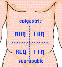
- Use the appropriate terminology to locate your findings:
- Right Upper Quadrant (RUQ)
- Right Lower Quadrant (RLQ)
- Left Upper Quadrant (LUQ)
- Left Lower Quadrant (LLQ)
- Midline:
- Epigastric
- Periumbilical
- Suprapubic
- Disorders in the chest will often manifest with abdominal symptoms. It is always wise to
examine the chest when evaluating an abdominal complaint.
- Consider the inguinal/rectal examination in males. Consider the pelvic/rectal
examination in females.
- Look for scars, striae, hernias, vascular changes, lesions, or rashes. [p336] [1]
- Look for movement associated with peristalsis or pulsations.
- Note the abdominal contour. Is it flat, scaphoid, or protuberant?
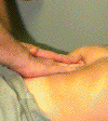
- Place the diaphragm of your stethoscope lightly on the abdomen. [p337] [2]
- Listen for bowel sounds. Are they normal, increased, decreased, or absent?
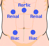
- Listen for bruits over the renal arteries, iliac arteries, and aorta.
- Percuss in all four quadrants using proper technique.
[p338]
- Categorize what you hear as tympanitic or dull. Tympany is normally present over most of
the abdomen in the supine position. Unusual dullness may be a clue to an underlying
abdominal mass.
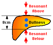
- Percuss downward from the chest in the right midclavicular line
until you detect the top edge of liver dullness.
- Percuss upward from the abdomen in the same line until you detect the
bottom edge of liver dullness.
- Measure the liver span between these two points. This measurement should be 6-12 cm in a
normal adult. [p341]
- Percuss the lowest costal interspace in the left anterior axillary line.
This area is normally tympanitic.
- Ask the patient to take a deep breath and percuss this area again. Dullness in this area
is a sign of splenic enlargement. [p345]
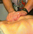
- Begin with light palpation. At this point you are mostly looking for
areas of tenderness. The most sensitive indicator of tenderness is the patient's facial
expression (so watch the patient's face, not your hands). Voluntary or involuntary
guarding may also be present. [p339]
- Proceed to deep palpation after surveying the abdomen lightly. Try to
identify abdominal masses or areas of deep tenderness.
- Place your fingers just below the right costal margin and press firmly.
- Ask the patient to take a deep breath.
- You may feel the edge of the liver press against your fingers. Or it may slide under
your hand as the patient exhales. A normal liver is not tender. [p342]
This method is useful when the patient is obese or when the examiner is small compared
to the patient.
- Stand by the patient's chest.
- "Hook" your fingers just below the costal margin and press firmly.
- Ask the patient to take a deep breath.
- You may feel the edge of the liver press against your fingers. [p344]
- Press down deeply in the midline above the umbilicus. ++
- The aortic pulsation is easily felt on most individuals.
- A well defined, pulsatile mass, greater than 3 cm across, suggests an aortic aneurysm.
[p350]
- Use your left hand to lift the lower rib cage and flank. ++
- Press down just below the left costal margin with your right hand.
- Ask the patient to take a deep breath.
- The spleen is not normally palpable on most individuals. [p346]
This is a test for peritoneal irritation. [p340] ++
- Warn the patient what you are about to do.
- Press deeply on the abdomen with your hand.
- After a moment, quickly release pressure.
- If it hurts more when you release, the patient has rebound tenderness. [4]
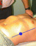
CVA tenderness is often associated with renal disease. [p349] ++
- Warn the patient what you are about to do.
- Have the patient sit up on the exam table.
- Use the heel of your closed fist to strike the patient firmly over the costovertebral
angles.
- Compare the left and right sides.
This is a test for peritoneal fluid (ascites). [p351] ++
- Percuss the patient's abdomen to outline areas of dullness and tympany.
- Have the patient roll away from you.
- Percuss and again outline areas of dullness and tympany. If the dullness has shifted to
areas of prior tympany, the patient may have excess peritoneal fluid. [5]
This is a test for appendicitis. [p353] ++
- Place your hand above the patient's right knee.
- Ask the patient to flex the right hip against resistance.
- Increased abdominal pain indicates a positive psoas sign.
This is a test for appendicitis. [p353] ++
- Raise the patient's right leg with the knee flexed.
- Rotate the leg internally at the hip.
- Increased abdominal pain indicates a positive obturator sign.
- Page numbers refer to Barbara Bates' A Guide to Physical
Examination and History Taking, Sixth Edition , published by Lippincott in 1995.
- Auscultation should be done prior to percussion and
palpation since bowel sounds may change with manipulation. Since bowel sounds are
transmitted widely in the abdomen, auscultation of more than one quadrant is not usually
necessary. If you hear them, they are present, period.
- Additional Testing - Tests marked with (++) may be skipped unless an
abnormality is suspected.
- Tenderness felt in the RLQ when palpation is performed on the left
is called Rovsing's Sign and suggests appendicitis. Rebound tenderness referred from the
left to the RLQ also suggests this disorder.
- Small amounts of peritoneal fluid are not usually detectable on
physical exam.
Author:
Richard Rathe, MD
Copyright: 1996-98 by the University of Florida