Cardiovascular Examination
- A Double-Headed, Double-Lumen Stethoscope
- A Blood Pressure Cuff
- A Moveable Light Source or Pen Light
- The patient must be properly undressed and in a gown for this
examination.
- The examination room must be quiet to perform adequate auscultation.
- Observe the patient for general signs of cardiovascular disease (finger clubbing,
cyanosis, edema, etc.).
- Compress the radial artery with your index and middle fingers. [p 274] [1]
- Note whether the pulse is regular or irregular. [p300]
- Count the pulse for 15 seconds and multiply by 4.
- Count for a full minute if the pulse is irregular. [2]
- Record the rate and rhythm.
| Pulse Classification in Adults (At Rest) |
| Normal |
Bradycardia |
Tachycardia |
| 60 to 100 bpm |
less than 60 bpm |
more than 100 |
| |
|
|
| Regular |
Regularly Irregular |
Irregularly Irregular |
| Evenly spaced beats, may vary slightly with respiration |
Regular pattern overall with "skipped" beats |
Chaotic, no real pattern, very difficult to measure rate accurately [2] |
[See below for children.]
- Observe for carotid pulsations. [p274]
- Place your fingers behind the patient's neck and compress the carotid artery on one side
with your thumb at or below the level of the cricoid cartilage. Press
firmly but not to the point of discomfort. [3]
- Assess the following:
- The amplitude of the pulse.
- The contour of the pulse wave.
- Variations in amplitude from beat to beat or with respiration.
- Repeat on the opposite side.
If the patient is middle aged or elderly, you should auscultate for bruits. A bruit is
often, but not always, a sign of arterial narrowing and risk of a stroke. [p276] ++ [4]
- Place the bell of the stethoscope over each carotid artery in turn. You
may use the diaphragm if the patient's neck is highly contoured.
- Ask the patient to stop breathing momentarily.
- Listen for a blowing or rushing sound--a bruit. Do not be confused by heart sounds or
murmurs transmitted from the chest.
The patient should not have eaten, smoked, taken caffeine, or engaged in vigorous
exercise within the last 30 minutes. The room should be quiet and the patient comfortable.
[p277]
- Position the patient's arm so the anticubital fold is level with the heart.
- Center the bladder of the cuff over the brachial artery approximately 2 cm above the
anticubital fold. Proper cuff size is essential to obtain an accurate
reading. Be sure the index line falls between the size marks when you apply the cuff.
Position the patient's arm so it is slightly flexed at the elbow.
- Palpate the radial pulse and inflate the cuff until the pulse disappears. This is a
rough estimate of the systolic pressure. [6]
- Place the stetescope over the brachial artery. [5]
- Inflate the cuff to 30 mmHg above the estimated systolic pressure.
- Release the pressure slowly, no greater than 5 mmHg per second.
- The level at which you consistantly hear beats is the systolic pressure. [7]
- Continue to lower the pressure until the sounds muffle and disappear. This is the
diastolic pressure. [8]
- Record the blood pressure as systolic over diastolic (120/70).
- Blood pressure should be taken in both arms on the first encounter. [9]
| Blood Pressure Classification in Adults |
| Category |
Systolic |
Diastolic |
| Normal |
<130 |
<85 |
| High Normal |
130-139 |
85-89 |
| Mild Hypertension |
140-159 |
90-99 |
| Moderate Hypertension |
160-179 |
100-109 |
| Severe Hypertension |
180-209 |
110-119 |
| Crisis Hypertension |
>210 |
>120 |
In children, pulse and blood pressure vary with the age. The
following table should serve as a rough guide:
| Average Pulse and Blood Pressure in Normal Children |
| Age |
Birth |
6mo |
1yr |
2yr |
6yr |
8yr |
10yr |
| Pulse |
140 |
130 |
115 |
110 |
103 |
100 |
95 |
| Systolic BP |
70 |
90 |
90 |
92 |
95 |
100 |
105 |
- Position the patient supine with the head of the table elevated 30 degrees. [p281] ++
- Use tangential, side lighting to observe for venous pulsations in the neck.

- Look for a rapid, double (sometimes triple) wave with each heart beat. Use light
pressure just above the sternal end of the clavicle to eliminate the pulsations and rule
out a carotid origin.
- Adjust the angle of table elevation to bring out the venous pulsation.

- Identify the highest point of pulsation. Using a horizontal line from this point,
measure vertically from the sternal angle. [10]
- This measurement should be less than 4 cm in a normal healthy adult.
- Position the patient supine with the head of the table slightly elevated. [p285]
- Always examine from the patient's right side.
- Inspect for precordial movement. Tangential lighting will make movements more visible.
- Palpate for precordial activity in general. You may feel "extras" such as
thrills or exaggerated ventricular impulses.
- Palpate for the point of maximal impulse (PMI or apical pulse). It is
normally located in the 4th or 5th intercostal space just medial to the midclavicular line
and is less than the size of a quarter.
- Note the location, size, and quality of the impulse.
- Position the patient supine with the head of the table slightly elevated. [p291]
- Always examine from the patient's right side. A quiet room is essential.
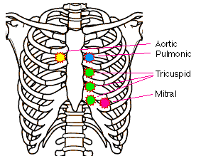
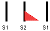
- Listen with the diaphragm at the right 2nd interspace near the sternum (aortic area).
- Listen with the diaphragm at the left 2nd interspace near the sternum (pulmonic area).
- Listen with the diaphragm at the left 3rd, 4th, and 5th interspaces near the sternum
(tricuspid area). [11]
- Listen with the diaphragm at the apex (PMI) (mitral area).
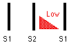
- Listen with the bell at the apex.
- Listen with the bell at the left 4th and 5th interspace near the
sternum. ++
- Have the patient roll on their left side. ++
- Listen with the bell at the apex.
- This position brings out S3 and mitral murmurs.
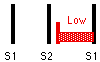
- Have the patient sit up, lean forward, and hold their breath in exhalation. ++
- Listen with the diaphragm at the left 3rd and 4th interspace near the sternum.
- This position brings out aortic murmurs.
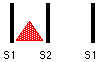
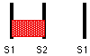
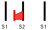
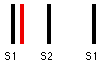
- Record S1, S2, (S3), (S4), as well as the grade and configuration of any murmurs
("two over six" or "2/6", "pansystolic" or
"crescendo").
| Murmurs and Extra Sounds |
Systolic Ejection
Innocent/Physiologic
Aortic/Pulmonic Stenosis |
Pansystolic
Mitral/Tricusp Regurgitation
|
Systolic Click
Late Systolic
Mitral Valve Prolapse
|
Early Diastolic
Aortic Regurgitation |
Mid Diastolic
Mitral/Tricusp Stenosis |
Opening Snap
Diastolic Rumble
Mitral Stenosis |
Ejection Sound
Aortic Valve Disease
|
S3
Normal in Children
Heart Failure |
S4
Physiologic
Various Diseases |
| Murmur Grades |
| Grade |
Volume |
Thrill |
| 1/6 |
very faint, only heard with optimal conditions |
no |
| 2/6 |
loud enough to be obvious |
no |
| 3/6 |
louder than grade 2 |
no |
| 4/6 |
louder than grade 3 |
yes |
| 5/6 |
heard with the stethoscope partially off the chest |
yes |
| 6/6 |
heard with the stethoscope completely off the chest |
yes |
- Page numbers refer to A Guide to Physical Examination and
History Taking, Sixth Edition by Barbara Bates, published by Lippincott in 1995.
- With an irregular pulse, the beats counted in any 30 second period
may not represent the overall rate. The longer you measure, the more these variations are
averaged out.
- Avoid compressing both sides a the same time. This could cut off the
blood supply to the brain and cause syncope. Avoid compressing the carotid sinus higher up
in the neck. This could lead to bradycardia and depressed blood pressure.
- Additional Testing - Tests marked with (++) may be skipped unless an
abnormality is suspected.
- Bell or Diaphragm? - Even though Korotkoff sounds are low frequency
and should be heard better with the bell, it is often difficult to apply the bell properly
to the anticubital fold. For this reason, it is common practice to use the diaphragm when
taking the blood pressure.
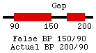
- Maximum Cuff Pressure - When the baseline blood pressure is already
known or hypertension is not suspected, it is acceptable in adults to inflate the cuff to
200 mmHg and go directly to auscultating the blood pressure. Be aware that there could be
an auscultory gap (a silent interval between the true systolic and diastolic pressures).
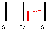
- Systolic Pressure - In situations where ausculation is not possible,
you can determine systolic blood pressure by palpation alone. Deflate the cuff until you
feel the radial or brachial pulse return. The pressure by auscultation would be
approximately 10 mmHg higher. Record the pressure indicating it was taken by palpation
(60/palp).
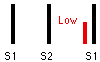
- Diastolic Pressure - If there is more than 10 mmHg difference
between the muffling and the disappearance of the sounds, record all three numbers
(120/80/45).
- Pressure Differences - If there is more than 10 mmHg difference
between the two arms, use the arm with the higher reading for subsequent measurements.
- Sternal Angle - The sternal angle is taken to be 5cm above the right
atrium. A jugular pulse 10cm above the sternal angle equates to a central venous pressure
of 15cm of water.
- Left Sternal Border - The left 3rd, 4th, and 5th interspaces are
considered the tricuspid area and are referred to as the Lower Left Sternal Border or
LLSB.
Author: Richard Rathe, MD
Copyright: 1996 by the University of Florida